Торакоскопия
На главную Написать письмо
История метода
Оборудование и инструменты
Диагностика и лечение заболеваний плевры
Торакоскопическая биопсия легкого
Лечение спонтанного пневмоторакса
Торакоскопическая диагностика и лечение заболеваний средостения
Резекция легкого при очаговом образовании
Анатомическая резекция легкого
Торакоскопические операции на пищеводе
Торакоскопическая симпатэктомия и спланхникэктомия
Лечение патологии грудной стенки
Лечение патологии диафрагмы
Консультации
Литература для специалистов
Лечение деформаций грудной клетки

Обратная связь


Главная страница >  Литература для специалистов >  Как не делать: Рестриктивная торакальная дистрофия после оперативного лечения воронкообразной деформации грудной клетки (pectus excavatum)

Как не делать: Рестриктивная торакальная дистрофия после оперативного лечения воронкообразной деформации грудной клетки (pectus excavatum)

Лечение деформаций грудной клетки
Роль видео-ассистированной торакальной хирургии в диагностике и лечении торакальных повреждений
Комбинированная видео-ассистированная медиастиноскопия и видео-ассистированая торакоскопия в лечении рака легкого
Таблица 1
Проспективное исследование: Систематическая диссекция лимфоузлов при раке легкого с помощью видео-ассистированной торакальной хирургии: Может ли она быть совершенной?
Таблица 1
Таблица 3
Таблица 1
Как не делать: Рестриктивная торакальная дистрофия после оперативного лечения воронкообразной деформации грудной клетки (pectus excavatum)
Биопсия легкого с использованием гармонического скальпеля: рандомизированное исследование одного института
Таблица 1
Таблица 2
Таблица 3
Видео-ассистированная фенестрация перикарда при фрагментрированных или рецидивных выпотах
Хронические последствия после торакоскопических операций по поводу доброкачественных заболеваний
Таблица 1
Солитарные фиброзные опухоли плевры: клинические характеристики, хирургическое лечение и исходы
Таблица 1 Критерии злокачественности 22 злокачественных опухолей
Таблица 2 Сравнение клинических и анатомических характеристик доброкачественных и злокачественных опухолей
Таблица 3 Патологические характеристики, хирургическое лечение и исходы в 3-х наблюдениях злокачественных рецидивных опухолях


Interactive Cardiovascular and Thoracic Surgery 3:566��(2004)
© 2004 European Association of Cardio-Thoracic Surgery

How not to do it: restrictive thoracic dystrophy after pectus excavatum repair
Francis Robicsek* and Alexander A. Fokin
Department of Thoracic and Cardiovascular Surgery, Carolinas Heart Institute, Carolinas Medial Center, 1000 Blythe Blvd., Charlotte, NC 28203, USA

* Corresponding author. Tel.: +1����? fax: +1����?. (E-mail: tjohn@sanger-clinic.com).

Received January 22, 2004; received in revised form June 8, 2004; accepted June 21, 2004


Abstract

The purpose of our paper is to call attention to acquired restrictive thoracic dystrophy (ARTD), an iatrogenic disease. The condition may occur following correction of pectus excavatum in young patients and is characterized by a reduced and restricted rib cage, usually with some recurrence of the deformity. The authors personal experience with five such patients includes analysis of their operative notes, radiographs, and long-term follow up. These, as well as literary, data indicate that the cause of ARTD is not that the operation is performed at an early age, but that inappropriate surgical technique was performed that may include radical chondrocostal resection, extirpation of growth centers, and suturing together of the perichondrial strips retrosternally, consequentially causing cartilaginous growth behind the sternum. A well-designed, conservative operation may be safely performed at any age, including in children less than 4 years old.


«On roentgenogram the entire rib-cage looked like that of a dog» Joel D. Cooper [1]

In the early 1980’s, attention was called to a group of patients who, after surgical repair of pectus excavatum, developed severe restrictive thoracic dystrophy. While the condition was coined by Haller as ‘acquired Jeune’s syndrome’ [2נ], because the components of Jeune’s syndrome extend beyond thoracic anatomy, [5] the nomer ‘acquired restrictive thoracic dystrophy’ (ARTD) appears to be more appropriate.

In addition to a history of pectus excavatum repair at an early age, the condition is characterized by a narrow torso and a small, immobile, ‘peaked’ anterior chest wall with horizontal ribs. ARTD patients are dyspneic to various degrees, with limited physical abilities. Conventional X-rays confirm the reduced size of the thorax and may also show some recurrence of the deformity. CT scans often demonstrate cartilaginous-osseous growth behind the sternum (Fig. 1). Pulmonary function studies reveal decreased vital capacity and forced expiratory volume [3,4,6]. While the exact mechanism of the ARTD is not entirely clear, it has been postulated that the development of this condition could be due to the fact that these patients were operated upon before 4 years of age [4,6]. In our opinion, based on personal observations of five patients, review of their medical records, as well as on literary data and court testimonies, ARTD is caused by faulty surgical technique that stops development and freezes the chest wall, which if this occurs in a young child, naturally could be devastating. It has been recognized that rib resection may cause chest deformity and scoliosis in young patients, especially when it is done extensively [3,4]. Several authors also emphasize the need to preserve the growth plates during surgery [4,6,7]. Experimental data also suggest that longitudinal growth of a normal rib occurs mainly at the sternal end [8].

РКТ Рестриктивной торакальной дистрофии при коррекции ВДГК
Fig. 1 CT scan of a patient with acquired restrictive thorax syndrome. Cartilaginous growth is seen behind the sternum.


Many ARTD patients were re-operated in an effort to improve their condition by enlarging their thoracic cage [4]. While these operations moderately expanded the thoracic cage, respiratory excursions of the thoracic wall remained restricted.
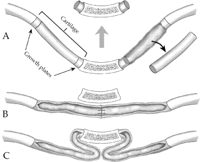
Our observations indicate that in patients with ARTD, the resection of the cartilages was too radical, both in number and extent. The records showed that the costal cartilages were extirpated from the sternum to the bony rib, from the second rib in most, and sometimes from the third rib down. This technique removes the growth plates of the cartilages, which are adjacent to the tip of the bony rib on one end, and at the sterno-chondral junction on the other (Fig. 2A).

Техника коррекции ВДГК по Robicsek
Fig. 2 Technical considerations for pectus deformity repair. (A) Resection of the deformed cartilages should not be too extensive and should leave the growth centers intact. (B) Perichondrial strips sutured together behind the sternum. (C) The excess length of the perichondrial strips may fold and unite under the sternum.


Analysis of CT scans in our group of patients revealed regrowth of ribs under the sternum. This may have occurred in one of the following ways: (1) The perichondrial strips from both sides were sutured together substernally to provide additional posterior support to the sternum (Fig. 2B); (2) After the deformity was corrected, the redundant perichondrial strips folded underneath the sternum (Fig. 2C). In either case, the perichondrium formed a cartilaginous-osseous plate, which, combined with loss of growth centers, left these children with a rib cage that did not move and did not grow.
While substernal suturing of ribs or the lower two perichondrial sheaths for sternal support was introduced in the 1970’s [9], Ravitch, in 1978, already expressed concern that such a maneuver may cause constriction of the lower chest [10].

All reported patients with ARTD were operated upon at less than 4 years old, thus evidently, age is an important factor [4]. Damage, however, may be inflicted by overzealous rib resections at any age. Naturally, if the growth of the anterior chest wall is brought to a halt in a very young child, the damage will be greater than if it would have occurred later in life. We and others reported repairs of pectus excavatum in patients under 3 years of age, with good long-term results [7,10], though some argue that there is no necessity to do surgery prior to 5ף or even 12 years of age [3]. We believe that it is all a matter of balancing the severity of the deformity and advantages of early correction versus postponing the surgery due to the possibility of complications. Our preference is to do the correction at preschool age, but we will not hesitate to operate at any age if the deformity is severe [7].

The occurrence of ARTD could be prevented by observing some simple safeguards: (A) The subperichondrial resection of the cartilages should be adequate, but not overzealous. The posterior perichondrium should be preserved to allow regeneration. Sleeve stitching of the costal perichondrium facilitates remodeling of the neocartilage. Resection of the second rib should be an exception and reserved for the most severe anomalies. (B) A short segment of the cartilage should be left in continuity with the bony rib, and also with the sternum. If the extent of the deformity requires the removal of the entire length of the cartilage or part of the bony rib, this should be limited to a few, certainly not to all ribs. (C) Perichondrial strips from both sides should not be sutured together behind the sternum. Small tissue defects in the lowermost part of the operative field may be left alone or corrected with rectus-fascioplasty.

The solution to the problem of preventing the development of acquired restrictive thoracic dystrophy after pectus excavatum repair is not to delay surgical intervention, but to do it appropriately.


2003 © www.thoracoscopy.ru
Design, programming, content
and promotion by A4-design
Rambler's Top100   Рейтинг@Mail.ru