 |

|
 |
|

|
Главная страница >
Литература для специалистов >
Комбинированная видео-ассистированная медиастиноскопия и видео-ассистированая торакоскопия в лечении рака легкого
Комбинированная видео-ассистированная медиастиноскопия и видео-ассистированая торакоскопия в лечении рака легкого
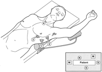
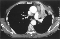
* Address reprint requests to Dr Mouroux, Service de Chirurgie Thoracique, Hôpital Pasteur, 30, Avenue de la Voie Romaine — BP 69, 06002 Nice Cedex 1, France Abstract Methods. Ten consecutive patients with lung neoplasms were evaluated.Indications for this combined approach included inconclusivefindings from imaging techniques concerning locoregional extensionand resectability; possible involvement of different structuresnot accessible to a single procedure; and failure to obtainhistologic diagnosis by a single technique. Results. Histologic diagnosis was obtained in 6 patients withoutpreoperative histologic typing. In 3 patients, in contrast withpreoperative imaging studies, combined thoracoscopy and mediastinoscopyshowed the resectability of the primary tumor and the absenceof metastatic mediastinal lymph nodes. These findings were confirmedat thoracotomy. In 3 other patients prevascular lymph nodesmetastases were found. They underwent neoadjuvant chemotherapy;at subsequent operation, a complete resection was possible.In the remaining four cases combined exploration proved definitivecontraindications for operation (recognition of oat-cell carcinoma,n = 2; T4 status, n = 1; T3N2, n = 1). Conclusions. Combined video-assisted mediastinoscopy and video-assistedthoracoscopy seems to be a safe and useful tool in the managementof selected patients with lung neoplasms. Both the extent ofprimary tumor and the possible intrathoracic spread may be exhaustivelyevaluated. In patients with left lung cancer a complete explorationof the aortopulmonary window is possible. Introduction Clinical staging is based on chest roentgenogram, bronchoscopy,computed tomography (CT) and, in some instances, magnetic resonanceimaging. Although generally useful, these methods are sometimesinadequate to provide correct information about locoregionalextension and resectability, especially in tumors involvinghilar regions [5,6]. Furthermore, they lack diagnostic accuracyin evaluation of mediastinal node metastases [5, 6]. Positronemission tomography (PET) seems a promising technique in thisfield [7]; however, its use will be probably limited to a fewinstitutions in the next years (In France, currently only 2centers are equipped with PET). Cervical mediastinoscopy, anterior mediastinotomy or mediastinoscopy,and video-assisted thoracoscopy (VT) have been widely used forboth diagnostic and staging purposes. Each of these techniqueshas evident merits in addition to limitations and «blind spots»[8]. Therefore, exact prethoracotomy staging of lung cancerremains difficult. This also explains the difficulties in selectionof patients for neoadjuvant treatment trials, in which histologicevidence of N2 status is almost always considered mandatory. In the present study we aimed at evaluating the clinical usefulnessof combined video-assisted mediastinoscopy (VM) and VT in themanagement of patients with lung neoplasms, with the idea thatthe combination of the two techniques would overcome their respectivelimitations, while preserving their individual merits. Material and methods Preoperative evaluation included chest roentgenogram, fiberopticbronchoscopy, and thoracic and upper abdominal CT scan by athird-generation apparatus making serial cuts 8 mm thick. BrainCT and bone scintigraphy were performed only in patients withclinically suspected cerebral or bone metastases. Nodal metastaseswere suspected in the presence of enlarged (short axis longerthan 1 cm) nodes at CT scan. Locations of the primary tumor were the left upper lobe in 6cases, the right upper lobe in 3 cases, and the left lower lobein 1 case. Histology was known preoperatively in 4 patients(squamous cell carcinoma in all cases). Informed consent was obtained by all the patients and the studywas conducted according to principles stated in the Declarationof Helsinki. Techniques  Video-assisted mediastinoscopy Video-assisted mediastinoscopy was carried out by using a specificallydesigned rigid scope, measuring 19 cm in length (Dahan/Lindermediastinoscope, model 8783.401, Richard Wolf, Knittlingen,Germany). The scope may be considered as a speculum: the inferiorvalve may be opened thus allowing optimal exposure of mediastinalstructures. The video-mediastinoscope is equipped with a distalfiberoptic lighting system. A mono CCD video camera (model INH002756 Karl Storz-Endoskope, Tuttlingen, Germany) was fittedto allow all the members of the surgical team to view. Surgical technique up to the introduction of the scope was essentiallythe same as that used for standard mediastinoscopy. After theparatracheal fascia opening and finger blunt dissection alongthe trachea, the video-mediastinoscope was inserted; its inferiorvalve was blocked in the open position. Further dissection wasperformed under direct visual control. The video-mediastinoscopewas handled by the assistant, thus allowing the surgeon to operatewith both hands. Generally, a metal blunt-tipped coagulation-suctiondevice and an endoscopic swab (Peanut, Auto Suture, Elancourt,France) or grasp was used simultaneously for dissection of anatomicalstructures. Trachea, superior vena cava, azygos vein, rightmain pulmonary artery, and left recurrent nerve were easilyidentified. Progression behind the right main pulmonary arteryallowed exploration of the two main bronchi and subcarinal area.Lymph nodes of levels 2, 4, and 7 were accessible for dissectionand biopsy. The possibility of operating with two instrumentsin his or her hands (possibly one to grasp and tract, the otherto dissect or coagulate) allowed the surgeon to completely enucleatelymph nodes in several instances. If a neoplastic infiltrationof the outer surfaces of trachea or main bronchi was suspected,a biopsy of these structures was carried out. Needle (18-gauge)puncture was performed before biopsy if doubts existed concerningthe possible vascular nature of a structure. Biopsies of enlargedlymph nodes were carried out; systematic biopsies of all theaccessible sites were also performed. Specimens were sent tothe laboratory for frozen sections. Minor hemorrhage occurringduring the operative maneuvers was controlled by coagulationor compression with a gauze. In some instances clips were usedto control minor bleeding or lymphatic leakage. The mediastinalbed was not drained routinely; if judged necessary, a 9-Ch (3mm) drainage attached to a suction device was used. Video-assisted thoracoscopy The possible extension of the primary tumors to mediastinalstructures could be evaluated easily. Lung could be retractedby endoscopic retractors; if necessary, mediastinal pleura orpericardium, or both, could be opened. Whenever safe, biopsieswith frozen sections were taken to confirm the neoplastic involvementof a structure or organ. Biopsies of lymph nodes nonaccessibleat cervical mediastinoscopy (those located in the anterior orthe inferior mediastinum) could also be obtained. Care was necessarywhen dissecting nodes in the aortopulmonary window to avoidinjury to vascular or nervous structures. Hemostasis and lymphostasisrequired accuracy and were achieved with the use of electrocoagulationor endoscopic clips. Generally a single drain was sufficientto adequately drain the thoracic cavity, especially if no injuryto lung parenchyma had been provoked. At combined VM and VT exploration, simultaneous instrumentalpalpation and transhillumination of hilar and mediastinal structureswas possible. On the left side, simultaneous dissection throughVM and VT allowed an exhaustive exploration of the aortopulmonarywindow: when there was no bulky tumor, all the anatomical structureswere recognized and dissection advanced up to the jointing ofmediastinoscopic and thoracoscopic instruments. All the encounteredlymph nodes (including the Botallo’s ligament nodes) couldbe sampled. If combined VM and VT demonstrated the resectability of thetumor, lung resection was performed immediately through a thoracotomy;otherwise the combined exploration was generally performed withan overnight stay. Results In 3 patients, in contrast with preoperative imaging studies,mediastinal exploration by combined VT and VM showed the immediateresectability of the primary tumor and no mediastinal lymphnode metastasis. In particular, in 1 patient the presence ofsuperior vena cava invasion and intrapulmonary metastatic spreadwas ruled out by VT with wedge resection of a suspected nodule.In the other 2 patients, involvement of proximal pulmonary arteryand tracheobronchial angle was ruled out. These 3 patients underwentimmediate lung resection with mediastinal nodal dissection (casenumbers 1 to 3, Table 1, Fig 2). The resection was completein all the instances. Pathologic examination of operative specimensconfirmed that the patients had been correctly staged by combinedVM and VT.  In 3 other patients with left lung cancer (case numbers 4 to6) prevascular nodal metastases were recognized at VT; in allthe cases VM showed no involvement of paratracheal and subcarinalnodes. Furthermore the tracheobronchial angle did not appearinvolved (a doubt existed on imaging studies in one case). In1 patient, the malignant nature of a concurrent pleural effusionwas ruled out by VT. All 3 patients received neoadjuvant chemotherapyand underwent subsequent operation. The resection was completeand pathologic examination showed no viable tumoral cells inprevascular nodes, but signs of necrosis, probably as the consequenceof chemotherapy. In 2 other patients (case numbers 7 and 8) a diagnosis of oat-cellcarcinoma was established on the basis of biopsy samples takenat VT. In patient 7, biopsy of a level 6 node showed tumoralinfiltration, whereas in patient 8 a hilar tumor was sampledand histologic diagnosis obtained. Biopsy samples of paratrachealand subcarinal stations taken at VM were negative, despite enlargeddimensions of lymph nodes. In both cases results from preoperativefiberoptic bronchoscopy with biopsy and CT guided fine-needleaspiration biopsy had been negative. These 2 patients underwentsubsequent chemotherapy. In another patient (number 9), preoperative imaging studieshad shown the presence of a right hilar tumor with paratrachealadenomegalies and pleural effusion. Repeated thoracocenteseshad been nondiagnostic. Video-assisted mediastinoscopy showedthe absence of retrovascular nodal metastasis, but a pleuralcarcinosis was found at thoracoscopy. He underwent systemicchemotherapy. In the last patient (number 10), thoracic CT scan had showna pleural effusion, paratracheal mediastinal adenopathies, andpossible involvement of chest wall. Preoperative thoracocentesishad not retrieved malignant cells. Frozen sections of biopsysamples of paratracheal nodes taken at VM showed malignant cells.At VT pleura was normal, but the presence of chest wall involvementwas confirmed. Definitive pathologic results were consistentwith a diagnosis of lung leiomyosarcoma. The patient was judgednot operable and chemotherapy was started. Comment Anterior mediastinoscopy or mediastinotomy are also used toapproach and sample prevascular mediastinal nodes, especiallyin patients with left upper lobe cancer [15, 16. Both presentthe advantage of technical simplicity, but their field of examinationis limited. Furthermore, it has been pointed out that in thesetting of staging left upper lobe tumors, biopsy through theanterior mediastinotomy is hazardous and assessment of mediastinumis limited to a digital examination of the aortic arch and subaorticfossa. Video-assisted thoracoscopy has been used for both diagnosticand staging purposes [18 — 20]. Indications for VT includesuspicion of pleural dissemination or intrapulmonary metastases. Video-assisted thoracoscopy has also been proposed toconfirm the presence of other T4 tumors (involving the aorta,atrium, esophagus, superior vena cava, spine) before enrollingpatients in protocols of neoadjuvant treatments [21]. In theleft hemithorax, prevascular (Naruke levels 5 and 6), inferiormediastinal (levels 8 and 9), subcarinal (level 7), and hilarnodes (level 10) are accessible to VT; in the right hemithorax,examination of all stations is possible [22]. Thus VT permitsexploration and biopsy of stations inaccessible by cervicalmediastinoscopy. Though in the right side VT would allow explorationof levels generally examined by cervical mediastinoscopy, thislast remains the «gold standard» for evaluation of retrovascularsuperior mediastinum, because of its proven characteristicsof accuracy and safety [22]. For this reason in this serieswe performed a VM in some patients with enlarged nodes althoughan ipsilateral VT was carried out. In this preliminary study we found that combined VM and VT wasa safe method requiring a short hospital stay. Operative timewas relatively short and will probably decrease in the futureas the learning curve reaches a plateau. In our hospital theService of Pathology is located in a separate building, thusjustifying the relatively long time for obtaining results fromfrozen sections, with a subsequent increase in operative time.We did not observe mortality or major morbidity related to combinedVM and VT. No port site seeding was observed at follow-up; itis noteworthy that this kind of late complication occurs rarelyafter VT for lung cancer [23]. Both T and N status could be adequately evaluated by combinedVM and VT. Based on preoperative imaging studies, a doubt concerninga possible involvement of mediastinal structures (superior venacava, aorta, proximal pulmonary artery, tracheobronchial angle)by the primary tumors existed in 5 patients; it was confirmedin 1 patient, but ruled out in the remaining 4 patients by thecombined exploration. The reliability of this evaluation wasconfirmed in the 3 patients who underwent immediate lung resectionand mediastinal nodal dissection. These 3 patients had beenconsidered N2 at clinical staging, but were classed N less than2 at combined mediastinal exploration. This finding was confirmedat pathologic examination of resected specimens. Three other patients underwent neoadjuvant chemotherapy forthe concurrent N2 status (prevascular nodes). They underwentsubsequent thoracotomy and a complete resection was possible.In the remaining 4 patients, the exploration by combined VMand VT allowed us to obtain a histologic diagnosis and a correctstaging, thus helping to guide subsequent medical treatment.Thus, in all the examined patients the combined explorationwas useful for clinical decision making. In our experience mediastinoscopy has been carried out usingthe video-assisted technique. Video-assisted mediastinoscopyhas been used for operating procedures, such as the Abruzzinioperation [24]. We have recently reported that VM is usefulin the staging of lung cancer [25]; although in that preliminarystudy no formal comparison was made between VM and standardmediastinoscopy, we remarked that VM offers better visualizationand allows the surgeon to operate with both hands, thus facilitatingdissection, biopsy, and hemostasis. However, VM is slightlymore expensive (+20%) than standard mediastinoscopy. As stated in the Material and Methods section, VM and VT canbe performed subsequently or simultaneously. The simultaneousapproach offers the advantages of avoiding changes in the patient’spositioning with an obvious shortening of operative times, atthe price of moderately increased difficulties in surgical techniques.The particular patient positioning is favorable for explorationof the anterior mediastinum; anterior retraction of the lungallows a complete exploration of posterior mediastinum as well.Furthermore, simultaneous instrumental palpation as well astranshillumination of hilar and mediastinal structures is possiblewith combined VM and VT. On the left side, simultaneous dissectionthrough VM and VT allows an exhaustive exploration of the aortopulmonarywindow. Endoscopic ultrasound probes are available for use during bothmediastinoscopy and VT; their use for the staging of lung cancerin selected cases has been proposed [26, 27]. In our institutionswe are currently developing a methodology for mediastinal ultrasoundexploration through combined VM and VT. This series evaluated the role of combined VM and VT in diagnosisand staging of lung cancer. Only one other case report discussedthe use of combined VT and standard mediastinoscopy in the stagingof a patient with left upper lobe carcinoma [28]. However, thecombination of standard cervical mediastinoscopy with anteriormediastinotomy has been evaluated extensively [17, 29, 30].In patients with left upper lobe tumors this technique is usedboth to examine the paratracheal nodes and to rule out directtumor invasion or fixed nodes around the aorta or main pulmonaryartery [17, 29]. Schreinemakers and colleagues [30] found thatperiaortic node metastases were present in 22.6% of patientswith a normal cervical mediastinoscopy and a very high resectabilitywas achieved in those subjects with negative parasternal andcervical examinations. Jiao and coworkers emphasized thevalue of bidigital examination through the anterior and thecervical incisions. However, in patients treated by subsequentlung resection with routine mediastinal node dissection, theyfound a high incidence (37.9%) of N2 disease unrecognized atmediastinal exploration through combined mediastinoscopy andmediastinotomy [17], thus suggesting that such kind of combinedmediastinal exploration is not optimal. Exploration throughcombined VM and VT offers probably better visualization, thuspermitting us to hypothesize that an increased sensitivity maybe achieved. Prospective studies are obviously mandatory tocompare these two types of combined approaches for mediastinalexploration. We think that when (a) imaging techniques provide inconclusiveinformation about resectability, (b) a doubt exists concerninga possible involvement of different structures not accessibleby a single procedure, or (c) a single procedure fails to providean histologic diagnosis, combined VM and VT represents a potenttool for diagnostic and staging purposes.
|
|
and promotion by A4-design |
|