Минимально-инвазивная эндоскопическая коррекция воронкообразной деформации грудной клетки (pectus excavatum)
Eur J Cardiothorac Surg 2002;21:869��
Minimally invasive endoscopic repair of pectus excavatum
Jeffrey P. Jacobsa*, James A. Quintessenzaa, Victor O. Morella, Luis M. Boteroa, Hugh M. van Geldera, Christo I. Tchervenkovb
a Division of Thoracic and Cardiovascular Surgery, All Children’s Hospital/University of South Florida College of Medicine, Cardiac Surgical Associates, 603 Seventh Street South, Suite 450, St. Petersburg, FL 33701, USA
b Division of Pediatric Cardiac Surgery, Montreal Children’s Hospital, Montreal, Canada
Received 29 October 2001; received in revised form 25 December 2001; accepted 22 January 2002.
* Corresponding author. Tel.: +1����? fax: +1����?
e-mail: jeffjacobs@msn.com
Abstract
Objective: We report our initial 3 years 4 months’ single institution experience in 31 consecutive patients with pectus excavatum treated with minimally invasive endoscopic pectus excavatum repair utilizing a modification of the ‘Nuss’ technique. Methods: Under general anesthesia, a curved steel bar is individually shaped for each patient to match the ideal chest wall shape and is placed through an endoscopically created retrosternal tunnel between two bilateral midaxillary line 2-cm incisions. The tunnels initially go along the outside of the rib cage, under the pectoral muscles. At the level of the sternum, these tunnels go retrosternal and communicate with each other. The steel bar is passed with the convexity facing posteriorly, within a protective flat silastic drain. Under endoscopic guidance, the curved steel bar is passed through one tunnel, under the sternum, and out the other tunnel. Once positioned, the bar is turned over, thereby correcting the deformity. An epidural catheter provides perioperative pain relief. Results: Minimally invasive endoscopic pectus excavatum repair has been performed on 31 patients (age: range 4.4㪷.0 years, median 15.0 years, mean 14.5 years). Median hospital length of stay is 4 days (range 3㪢 days, mean 4.6 days). Pneumothorax occurred in five patients requiring tube thoracostomy in three. One patient developed delayed bilateral pleural effusions requiring drainage. Two patients developed evidence of sterile seroma formation at the skin incision several months after minimally invasive repair of pectus excavatum. These seromas resolved with non-interventional conservative medical treatment. No other complications occurred. Conclusion: The minimally invasive endoscopic pectus repair is safe and effective and currently our procedure of choice for primary pectus excavatum in all ages. Endoscopic visualization facilitates the safe creation of the retrosternal tunnel. Short-term results have been excellent. Further follow-up will be necessary to determine long-term results.
Key Words: Pectus excavatum • Nuss technique
1. Introduction
Pectus excavatum, a relatively common congenital chest wall deformity in children, is a depression of the sternum that commonly starts at the angle of Louis, and is deepest at the xiphisternal junction [1]. When severe, pectus excavatum deformity can cause cardiopulmonary insufficiency from the compression of the right atrium and right ventricle and diminished vital capacity of the lungs [1].
A variety of techniques are available to repair pectus excavatum deformity (Table 1). The classic surgical repair of pectus excavatum is the Ravitch repair, which entails subperichondrial resection of the deformed cartilages and a sternal osteotomy [2]. Retrosternal support with a metal bar is often used with this approach [3ס]. Outcome with a temporary sternal bar is felt by some surgical teams to be superior to outcome without a bar [3]. Other pectus excavatum repair techniques include the sternal turnover technique, the unilateral costoplasty technique, and the reconstruction with silicone implants.
Table 1. Techniques of pectus excavatum repaira
In 1987, Dr. Donald Nuss developed a minimally invasive technique for treatment of pectus excavatum using a convex steel bar placed beneath the pectus deformity and turned to correct the defect [6]. Excellent long-term results with this technique applied to 42 patients under age 15 were published in 1998 [6]. Questions regarding the safety of this technique have been raised [3].
In an effort to address these safety concerns, at All Children’s Hospital, we have made two modifications of Dr. Nuss’ technique. Our modifications of the Nuss procedure are designed to increase the surgeon’s control during the creation of the retrosternal tunnel and the passage of the steel retrosternal bar. We report our initial 3 years 4 months’ single institution experience in 31 consecutive patients with pectus excavatum treated with minimally invasive endoscopic pectus excavatum repair utilizing this modification of the ‘Nuss’ technique.
2. Materials and methods
2.1. Surgical techniques
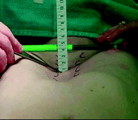
Under general anesthesia, an epidural catheter is placed to aid in perioperative pain control. A sterile marking pen marks the skin at the interspaces of maximal pectus depth on each side of the sternum. The preoperative anatomy is documented with photography in the anteroposterior plane (Fig. 1) and lateral planes.
Fig. 1. The preoperative anatomy is documented with photography in the anteroposterior plane. A sterile marking pen marks the skin at the interspaces of maximal pectus depth on each side of the sternum. In this example, the pectus depth measures 4.5 cm. Eventually, a retrosternal stainless steel bar will be placed from midaxillary line to midaxillary line at the level of maximal pectus depth. The length from midaxillary line to midaxillary line will be measured in order to size for the proper bar size.
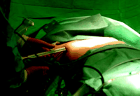
At the level of maximum pectus depth where sternal bone is present, the length from midaxillary line to midaxillary line is measured in order to select the proper size template and bar. A curved steel bar is individually shaped for each patient to match the ideal chest wall shape by curving the bar to match the curvature of a template fitted on the patient’s chest. Two small 2-cm thoracic incisions are made in the midaxillary line bilaterally, at the level of maximum pectus depth where sternal bone is present. These incisions are matured to the chest wall. Clamps ranging from small to large will be utilized to create a retrosternal tunnel, which will be deep to the pectoralis musculature but superficial to the ribs and intercostal musculature. Successively larger clamps will mature the tunnel (Fig. 2).
Fig. 2. This figure demonstrates the endoscopic tunneling device and the clamps used for tunneling. Clamps ranging from small to large will be utilized to create the retrosternal tunnel. This tunnel will be deep to the pectoralis musculature but superficial to the ribs and intercostal musculature. Successively larger clamps will mature the tunnel.
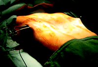
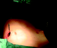
Two tunnels are fashioned bilaterally along the outside of the rib cage, under the pectoral muscles (Fig. 3). At the level of the sternum, these tunnels go retrosternal and communicate with each other (Fig. 4). The curved steel bar is passed through one tunnel, under the sternum, and out the other tunnel. The steel bar is inserted with the convexity facing posteriorly (Fig. 5), and when it is in position, the bar is turned over, thereby correcting the deformity (Fig. 6). The bar is then secured in position with sutures bilaterally. For larger patients with larger defects, two bars may be used. Occasionally, a small steel, grooved plate is used to stabilize the bar(s).
Fig. 3. Once the tunnel reaches the sternum, the endoscopic tunneling device is utilized to visualize the clamp entering the interspace of maximal pectus depth and passing retrosternal. This endoscopic tunneler is the same instrument used in our adult cardiac program for endoscopic saphenous vein procurement. The endoscopic view of the tunnel allows visualization of the safe passage of the clamp under the sternum and anterior to the heart. The mediastinum and pericardium can be seen pulsating posterior to the clamp.
Fig. 4. The larger curved clamp now passes under the sternum easily. The clamp now passes in one midaxillary skin incision and out the other midaxillary skin incision.
Fig. 5. The bar is now within the tunnel and in a retrosternal position. Next, the bar edges will be flared inward bilaterally. Then the bar will be rotated, correcting the pectus excavatum deformity.
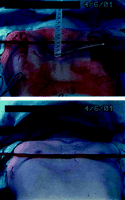
Fig. 6. This figure demonstrates the preoperative and postoperative appearance of the chest wall.
At All Children’s Hospital, we have made two modifications of Dr. Nuss’ technique in an effort to increase the surgeon’s control during the creation of the retrosternal tunnel and the passage of the steel retrosternal bar. First, we utilize the endoscopic saphenous vein harvesting equipment from our adult cardiac program to endoscopically create the tunnels under visual guidance (Figs. 2 and 3). We have found tremendous similarity between the tunnel necessary for endoscopic saphenous vein harvesting and the tunnels for minimally invasive pectus repair and we feel that the improved visualization afforded by this technique might decrease the potential for cardiac or mammary artery injury during minimally invasive pectus repair. Second, once the tunnel is created, we then use a clamp to pull two heavy sutures through the tunnel; one of these sutures acts as a backup and the second suture pulls a flat silastic mediastinal drain through the tunnel. The steel bar is then passed through the retrosternal space within the flat silastic mediastinal drain, thereby protecting surrounding structures.
When permanent chest wall remolding has occurred, bar removal is performed as an outpatient procedure. We currently recommend bar removal 2 years after insertion in patients under age 10 years at the time of bar insertion. For patients aged between 10 and 13 at the time of bar insertion, we recommend bar removal 3 years after insertion. For patients aged over 13 at the time of bar insertion, we recommend bar removal 4 years after insertion.
2.2. Patients
This series reviews the initial 31 consecutive patients undergoing minimally invasive endoscopic pectus excavatum repair at All Children’s Hospital (Saint Petersburg, FL, USA). Patient age ranged from 4.4 to 31.0 years (median 15.0 years, mean 14.5 years). Patient weight ranged from 17.8 to 120 kg (median 52.1 kg, mean 55.7 kg). No patients had associated structural cardiac anomalies although one patient had associated Wolff — Parkinson — White syndrome which was treated with transcatheter ablation prior to pectus repair. All patients had primary pectus excavatum deformity and evidence of associated physiological or psychological compromise. No patient had recurrent pectus excavatum deformity after prior attempted repair.
3. Results
Operative mortality was zero. No bleeding complications occurred. No injury or damage to underlying cardiac structures or the mammary arteries or veins occurred.
Epidural anesthesia provided excellent perioperative pain control in the initial 48㫠 h after the procedure. Median hospital length of stay was 4 days (range 3㪢 days, mean 4.6 days).
Pneumothorax occurred in five patients, requiring tube thoracostomy in three. We perform a chest radiograph prior to leaving the operating theater. Because no holes in the lung are created, we now feel that a small to moderate pneumothorax may be managed conservatively with supplemental oxygen therapy and without chest tube placement. These small to moderate pneumothoraces are well tolerated and can be observed to resolve with daily chest radiography.
One patient developed delayed bilateral pleural effusions requiring drainage. Two patients developed evidence of sterile seroma formation at the skin incision several months after minimally invasive repair of pectus excavatum. In one patient, this seroma formation was related to local trauma; the seroma developed spontaneously in the second patient. In both patients, the seromas resolved within 10 days with non-interventional conservative medical treatment (warm compresses and non-steroidal anti-inflammatory drugs). Neither patient required drainage of the seromas.
No other complications occurred. We have not had any wound infections or patients who required bar revision.
4. Discussion
Dr. Nuss has reported excellent results with the minimally invasive repair of pectus excavatum. In 1998, he reported his 10-year experience with 42 patients under 15 years old treated by the minimally invasive technique. Of 42 patients who had the minimally invasive procedure, 30 had undergone bar removal. Initial excellent results were maintained in 22, good results in four, fair in two, and poor in two, with mean follow-up since surgery of 4.6 years (range 1ץ.2 years). Average blood loss was 15 ml. Average length of hospital stay was 4.3 days. Patients returned to full activity after 1 month. Complications were pneumothorax in four patients, requiring thoracostomy in one patient; superficial wound infection in one patient; and displacement of the steel bar requiring revision in two patients. The fair and poor results occurred early in the series because (1) the bar was too soft (three patients), (2) the sternum was too soft in one of the patients with Marfan’s syndrome, and (3) in one patient with complex thoracic anomalies, the bar was removed too soon.
Since the first report by Dr. Nuss of the technique for minimally invasive repair of pectus excavatum, the popularity and demand for this operation has increased dramatically [7]. A survey reported in 2000 in the Journal of Pediatric Surgery reported that of 74 responders, 31 (42%) use minimally invasive repair of pectus excavatum as their procedure of choice. This survey report reviewed 251 cases of minimally invasive repair of pectus excavatum. Complications reported were bar displacement or rotation requiring reoperation (9.2%), pneumothorax requiring tube thoracostomy (4.8%), infectious complications (2%), pleural effusion (2%), thoracic outlet obstruction (0.8%), cardiac injury (0.4%), sternal erosion (0.4%), pericarditis (0.4%), and anterior thoracic artery pseudoaneurysm (0.4%). Three patients (1.2%) required early strut removal. Reoperation using the open modified Ravitch approach was performed in two patients (0.8%). Most surgeons indicated that teenaged patients (>15 years old) were at higher risk for complications. Overall patient satisfaction was rated as excellent or good (96.5%) [7].
Several publications have documented that the Nuss procedure leads to satisfactory results in the overwhelming majority of patients [6,7] but that complications may occur [3,6פ] and that a learning curve exists [7,8]. Nearly all reported complications are transient and easily treatable with minimal morbidity. The exception to this statement is the reported complication of cardiac injury [3,7]. Although this complication is unlikely, it is unacceptable. Our modifications of the ‘Nuss’ technique are designed to eliminate this complication. Endoscopic visualization during tunnel creation will allow for safer tunnel creation. The endoscopic saphenous vein harvesting equipment is ideal for this purpose. The learning curve is potentially less treacherous with this increased visualization. Furthermore, by passing the steel bar through the retrosternal space within the flat silastic mediastinal drain, the underlying cardiac structures are protected. These slight modifications of Dr. Nuss’ technique allow for increased control during the creation of the retrosternal tunnel and the passage of the bar.
Minimally invasive endoscopic repair of pectus excavatum is suitable for any patient with primary pectus excavatum deformity and evidence of associated physiological or psychological compromise. This technique is not suitable for recurrent pectus excavatum deformity after prior attempted repair. Although this technique may be applied to any patient age, we prefer to wait until the age of 4 years. While the best candidates are those patients with symmetrical chest wall deformities, patients with asymmetrical deformities also will benefit from this technique. We advise these patients with asymmetrical deformities (and their families) that a slight residual asymmetry may persist after repair. Most patients and families prefer this slight residual asymmetry to the larger scars associated with other conventional techniques of pectus excavatum repair. We have successfully used this technique on extremely severe and deep (6 cm) deformities with outstanding results. The minimally invasive endoscopic repair of pectus excavatum is safe and effective and currently our procedure of choice for primary pectus excavatum in all ages.
Endoscopic visualization during tunnel creation can help eliminate the dreaded complication of visceral perforation. To prevent bar migration, we secure the bar bilaterally to the chest wall fascia with heavy non-absorbable monofilament sutures. One posterior non-absorbable suture is passed through a hole in the bar. Two additional anterior non-absorbable sutures are passed around the bar. Using this technique, we have not seen the reported complication of bar migration or ‘wandering’ of the Nuss bar.
5. Conclusion
The minimally invasive endoscopic repair of pectus excavatum is safe and effective, and is currently our procedure of choice for primary pectus excavatum in all ages. Endoscopic visualization facilitates the safe creation of the retrosternal tunnel. Short-term results have been excellent. Further follow-up will be necessary to determine long-term results.
Footnotes
Presented at the joint 15th Annual Meeting of the European Association for Cardio-thoracic Surgery and the 9th Annual Meeting of the European Society of Thoracic Surgeons, Lisbon, Portugal, September 16㪫, 2001.
References
Backer C.L., Mavroudis C.:. The Society of Thoracic Surgeons Congenital Heart Surgery Nomenclature and Database Project: vascular rings, tracheal stenosis, and pectus excavatum. Ann Thorac Surg 2000;69(Suppl):S308.[Abstract/Free Full Text]
Ravitch M.M. The operative treatment of pectus excavatum. Ann Surg 1949;129:429��.
Willekes C.L., Backer C.L., Mavroudis C. A 26-year review of pectus deformity repairs, including simultaneous intracardiac repair. Ann Thorac Surg 1999;67(2):511��.[Abstract/Free Full Text]
Haller J.A., Scherer L.R., Turner C.S., Colombani P.M. Evolving management of pectus excavatum based on a single institutional experience of 664 patients. Ann Surg 1989;209:578��.[Medline]
Shamberger R.C., Welch K.J. Surgical repair of pectus excavatum. J Pediatr Surg 1988;23:615��.[Medline]
Nuss D., Kelly R.E., Jr, Croitoru D.P., Katz M.E. A 10-year review of a minimally invasive technique for the correction of pectus excavatum. J Pediatr Surg 1998;33(4):545��.[Medline]
Hebra A., Swoveland B., Egbert M., Tagge E.P., Georgeson K., Othersen H.B., Jr, Nuss D. Outcome analysis of minimally invasive repair of pectus excavatum: review of 251 cases. J Pediatr Surg 2000;35(2):252��.[Medline]
Engum S., Rescorla F., West K., Rouse T., Scherer L.R., Grosfeld J. Is the grass greener? Early results of the Nuss procedure. J Pediatr Surg 2000;35(2):246��.[Medline]
|


